
Engineers at Stanford have demonstrated a high-resolution endoscope that is as thin as a human hair with a resolution four times better than previous devices of similar design. The so-called micro-endoscope is a significant step forward in high-resolution, minimally invasive bio-imaging, with potential applications in research and clinical practice. Micro-endoscopy could enable new methods in diverse fields ranging from study of the brain to early cancer detection.
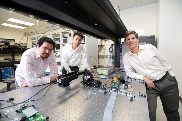
The new endoscope was developed by a team under the direction of Joseph Kahn, professor of electrical engineering at the Stanford School of Engineering. The results were published recently in the journal Optics Express and showcased in the Optical Society of America’s “Spotlight on Optics.”
Their prototype can resolve objects about 2.5 microns in size, and a resolution of 0.3 microns is easily within reach. A micron is one thousandth of a millimeter. By comparison, today’s high-resolution endoscopes can resolve objects only to about 10 microns. The naked eye can see objects down to about 125 microns.
Light paths
Kahn is best known for his work in fiberoptic communications – the ultra-fast data pipes essential to the Internet and large-scale data centers. His work on endoscopy began two years ago when he and a fellow Stanford electrical engineer, Olav Solgaard, were discussing biophotonics – a field of light-based technologies used in studying biological systems.
“Olav wanted to know if it would it be possible to send light through a single hair-thin fiber, form a bright spot inside the body and scan it to record images of living tissue,” said Kahn.
The opportunity and the challenge, Kahn and Solgaard knew, rested in multimode fibers in which light travels via many different paths, known in optics as modes; hence the name, multimode fiber. Light is very good at conveying complex information through such fibers – whether computer data or images – but it gets scrambled potentially beyond recognition along the way.
Kahn devised a way to undo the scrambling of information by using a miniature liquid crystal display called a spatial light modulator. To make this possible, Kahn and his graduate student Reza Nasiri Mahalati developed an adaptive algorithm – a specialized computer program – by which the spatial light modulator learned how to unscramble the light. Several years before, Kahn had set world records for transmission speeds using a similar trick to unscramble computer data transmitted through multimode fibers.
Research on the micro-endoscope took an unexpected and fortunate turn when Nasiri Mahalati mentioned seminal work in magnetic resonance imaging (MRI) done by John Pauly, another Stanford electrical engineer. Pauly had used random sampling to dramatically speed up image recording in MRIs.
“Nasiri Mahalati said, ‘Why not use random patterns of light to speed up imaging through multimode fiber?’ and that was it. We were on our way,” said Kahn. “The record-setting micro-endoscope was born.”